
Magnetic resonance elastography
Magnetic resonance elastography (MRE) is a form of elastography that specifically leverages MRI to quantify and subsequently map the mechanical properties (elasticity or stiffness) of soft tissue. First developed and described at Mayo Clinic by Muthupillai et al. in 1995, MRE has emerged as a powerful, non-invasive diagnostic tool, namely as an alternative to biopsy and serum tests for staging liver fibrosis.[1][2][3][4][5]
Diseased tissue (e.g. a breast tumor) is often stiffer than the surrounding normal (fibroglandular) tissue,[6] providing motivation to assess tissue stiffness.[7] This principle of operation is the basis for the longstanding practice of palpation, which, however, is limited (except at surgery) to superficial organs and pathologies, and by its subjective, qualitative nature, depending on the skill and touch sensitivity of the practitioner. Conventional imaging techniques of CT, MRI, US, and nuclear medicine are unable to offer any insight on the elastic modulus of soft tissue.[2] MRE, as a quantitative method of assessing tissue stiffness, provides reliable insight to visualize a variety of disease processes which affect tissue stiffness in the liver, brain, heart, pancreas, kidney, spleen, breast, uterus, prostate, and skeletal muscle.[2][3][8]
MRE is conducted in three steps: first, a mechanical vibrator is used on the surface of the patient's body to generate shear waves that travel into the patient's deeper tissues; second, an MRI acquisition sequence measures the propagation and velocity of the waves; and finally this information is processed by an inversion algorithm to quantitatively infer and map tissue stiffness in 3-D.[2][3] This stiffness map is called an elastogram, and is the final output of MRE, along with conventional 3-D MRI images as shown on the right.[2]
Mechanics of soft tissue[edit]
MRE quantitatively determines the stiffness of biological tissues by measuring its mechanical response to an external stress.[3] Specifically, MRE calculates the shear modulus of a tissue from its shear-wave displacement measurements.[7] The elastic modulus quantifies the stiffness of a material, or how well it resists elastic deformation as a force is applied. For elastic materials, strain is directly proportional to stress within an elastic region. The elastic modulus is seen as the proportionality constant between stress and strain within this region. Unlike purely elastic materials, biological tissues are viscoelastic, meaning that it has characteristics of both elastic solids and viscous liquids. Their mechanical responses depend on the magnitude of the applied stress as well as the strain rate. The stress-strain curve for a viscoelastic material exhibits hysteresis. The area of the hysteresis loop represents the amount of energy lost as heat when a viscoelastic material undergoes an applied stress and is distorted. For these materials, the elastic modulus is complex and can be separated into two components: a storage modulus and a loss modulus. The storage modulus expresses the contribution from elastic solid behavior while the loss modulus expresses the contribution from viscous liquid behavior. Conversely, elastic materials exhibit a pure solid response. When a force is applied, these materials elastically store and release energy, which does not result in energy loss in the form of heat.[9]
Yet, MRE and other elastography imaging techniques typically utilize a mechanical parameter estimation that assumes biological tissues to be linearly elastic and isotropic for simplicity purposes.[10] The effective shear modulus can be expressed with the following equation:

![{\displaystyle \mu =E/[2(1+\nu )]}](https://wikimedia.org/api/rest_v1/media/math/render/svg/43fa67a31086c574b65db86e0df38405731f2b95)
where is the elastic modulus of the material and is the Poisson's ratio.


The Poisson's ratio for soft tissues is approximated to equal 0.5, resulting in the ratio between the elastic modulus and shear modulus to equal 3.[11] This relationship can be used to estimate the stiffness of biological tissues based on the calculated shear modulus from shear-wave propagation measurements. A driver system produces and transmits acoustic waves set at a specific frequency (50–500 Hz) to the tissue sample. At these frequencies, the velocity of shear waves can be about 1–10 m/s.[12][13] The effective shear modulus can be calculated from the shear wave velocity with the following:[14]

where is the tissue density and is the shear wave velocity.


Recent studies have been focused on incorporating mechanical parameter estimations into post-processing inverse algorithms that account for the complex viscoelastic behavior of soft tissues. Creating new parameters could potentially increase the specificity of MRE measurements and diagnostic testing.[15][16]
Applications[edit]
Liver[edit]

Liver fibrosis is a common condition arising in many liver diseases. Progression of fibrosis can lead to cirrhosis and end-stage liver disease. MRE-based measurement of liver stiffness has emerged as the most accurate non-invasive technique for detecting and staging liver fibrosis. MRE provides quantitative maps of tissue stiffness over large regions of the liver. Abnormally increased liver stiffness is a direct consequence of liver fibrosis. The diagnostic performance of MRE in assessing liver fibrosis has been established in multiple studies.[17][18][16][19]
Liver MRE examinations are performed in MRI systems that have been equipped for the technique. Patients should fast for 3 to 4 hours prior to their MRE exam to allow for the most accurate measurement of liver stiffness.[20][21][22] Patients lie supine in the MRI scanner for the examination. A special device is placed on the right side of the chest wall over the liver to apply gentle vibration which generates propagating shear waves in the liver. Imaging is for MRE is very quick, with data acquired in a series of 1-4 periods of breath-holding, each lasting 15–20 seconds.
A standardized approach for performing and analyzing liver MRE exams has been documented by the RSNA Quantitative Imaging Biomarkers Alliance.[23] The technical success rate of Liver MRE is very high (95-100%)[24][25][26]
Brain[edit]
| Magnetic resonance elastography | |
|---|---|
 Magnetic resonance elastography of the brain. A T1 weighted anatomical image is shown in the top-left, and the corresponding T2 weighted image from the MRE data is shown in the bottom-left. The wave image used to make the elastogram is shown in the top-right, and the resulting elastogram is in the bottom-right. | |
| Purpose | measures the mechanical properties of soft tissues |
MRE of the brain [27] was first presented in the early 2000s.[28][29] Elastogram measures have been correlated with memory tasks,[30] fitness measures,[31] and progression of various neurodegenerative conditions.[27] For example, regional and global decreases in brain viscoelasticity have been observed in Alzheimer's disease[32][33] and multiple sclerosis.[34][35] It has been found that as the brain ages, it loses its viscoelastic integrity due to degeneration of neurons and oligodendrocytes.[36][37] A recent study looked into both the isotropic and anisotropic stiffness in brain and found a correlation between the two and with age, particularly in gray matter.[38]
MRE may also have applications for understanding the adolescent brain. Recently, it was found that adolescents have regional differences in brain viscoelasticity relative to adults.[39][40]
MRE has also been applied to functional neuroimaging. Whereas functional magnetic resonance imaging (fMRI) infers brain activity by detecting relatively slow changes in blood flow, functional MRE is capable of detecting neuromechanical changes in the brain related to neuronal activity occurring on the 100-millisecond scale.[41]
Kidney[edit]
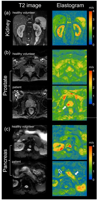
MRE has also been applied to investigate the biomechanical properties of the kidney. The feasibility of clinical renal MRE was first reported in 2011 for healthy volunteers [42] and in 2012 for renal transplant patients.[43] Renal MRE is more challenging than MRE of larger organs such as the brain or liver due to fine mechanical features in the renal cortex and medulla as well as the acoustically shielded position of the kidneys within the abdominal cavity. To overcome these challenges, researchers have been looking at different passive drivers and imaging techniques to best deliver shear waves to the kidneys.[44][45][46][47][48] Studies investigating renal diseases such as renal allograft dysfunction,[49][50][51][52] lupus nephritis,[53] immunoglobulin A nephropathy (IgAN),[54] diabetic nephrology,[55] renal tumors[56] and chronic kidney disease[57] demonstrate that kidney stiffness is sensitive to kidney function[58][59] and renal perfusion.[58][60]
Prostate[edit]
The prostate can also be examined by MRE, in particular for the detection and diagnosis of prostate cancer.[61] To ensure good shear wave penetration in the prostate gland, different actuator systems were designed and evaluated.[62][63] Preliminary results in patients with prostate cancer showed that changes in stiffness allowed differentiation of cancerous tissue from normal tissue.[64] Magnetic Resonance Elastography has been successfully used in patients with prostate cancer showing high specificity and sensitivity in differentiating prostate cancer from benign prostatic diseases [65][66] (see figure on right (b)). Even higher specificity of 95% for prostate cancer was achieved when Magnetic Resonance Elastography was combined with systematic image interpretation using PI-RADS (version 2.1).[66][67]
Pancreas[edit]
The pancreas is one of the softest tissues in the abdomen. Given that pancreatic diseases including pancreatitis and pancreatic cancer significantly increase stiffness, MRE is a promising tool for diagnosing benign and malignant conditions of the pancreas. Abnormally high pancreatic stiffness was detected by MRE in patients with both acute and chronic pancreatitis.[68] Pancreatic stiffness was also used to distinguish pancreatic malignancy from benign masses [69] and to predict the occurrence of pancreatic fistula after pancreaticoenteric anastomosis.[70] Quantification of the volume of pancreatic tumors based on tomoelastographic measurement of stiffness was found to be excellently correlated with tumor volumes estimated by contrast-enhanced computed tomography.[71] In patients with pancreatic ductal adenocarcinoma stiffness was found to be elevated in the tumor as well as in pancreatic parenchyma distal to the tumor, suggesting heterogeneous pancreatic involvement [72] (figure on right (c)).
See also[edit]
References[edit]
- ^ Hirsch, Sebastian; Braun, Jürgen; Sack, Ingolf (2016). Magnetic Resonance Elastography | Wiley Online Books. doi:10.1002/9783527696017. ISBN 9783527696017.
- ^ a b c d e Mariappan YK, Glaser KJ, Ehman RL (2010). "Magnetic resonance elastography: a review". Clin Anat. 23 (5): 497–511. doi:10.1002/ca.21006. PMC 3066083. PMID 20544947.
- ^ a b c d Glaser KJ, Manduca A, Ehman RL (14 September 2012). "Review of MR elastography applications and recent developments". J Magn Reson Imaging. 36 (4): 757–74. doi:10.1002/jmri.23597. PMC 3462370. PMID 22987755.
- ^ Chen J, Yin M, Glaser KJ, Talwalkar JA, Ehman RL (2013). "MR Elastography of Liver Disease: State of the Art". Appl Radiol. 42 (4): 5–12. doi:10.37549/AR1982. PMC 4564016. PMID 26366024.
- ^ Ingolf Sack: Magnetic resonance elastography from fundamental soft-tissue mechanics to diagnostic imaging. In: Nature Reviews Physics. 5, 2023, S. 25, doi:10.1038/s42254-022-00543-2.
- ^ Pepin KM, Ehman RL, McGee KP (2015). "Magnetic resonance elastography (MRE) in cancer: Technique, analysis, and applications". Prog Nucl Magn Reson Spectrosc. 90–91: 32–48. doi:10.1016/j.pnmrs.2015.06.001. PMC 4660259. PMID 26592944.
- ^ a b Muthupillai R, Lomas DJ, Rossman PJ, Greenleaf JF, Manduca A, Ehman RL (September 1995). "Magnetic resonance elastography by direct visualization of propagating acoustic strain waves". Science. 269 (5232): 1854–7. Bibcode:1995Sci...269.1854M. doi:10.1126/science.7569924. PMID 7569924.
- ^ Wang, Jin; Deng, Ying; Jondal, Danielle; Woodrum, David M.; Shi, Yu; Yin, Meng; Venkatesh, Sudhakar K. (2018). "New and Emerging Applications of Magnetic Resonance Elastography of Other Abdominal Organs". Topics in Magnetic Resonance Imaging. 27 (5): 335–352. doi:10.1097/RMR.0000000000000182. ISSN 0899-3459. PMC 7042709. PMID 30289829.
- ^ Wineman A (2009). "Nonlinear Viscoelastic Solids—A Review". Mathematics and Mechanics of Solids. 14 (3): 300–366. doi:10.1177/1081286509103660. ISSN 1081-2865. S2CID 121161691.
- ^ Mariappan YK, Glaser KJ, Ehman RL (July 2010). "Magnetic resonance elastography: a review". Clinical Anatomy. 23 (5): 497–511. doi:10.1002/ca.21006. PMC 3066083. PMID 20544947.
- ^ Low G, Kruse SA, Lomas DJ (January 2016). "General review of magnetic resonance elastography". World Journal of Radiology. 8 (1): 59–72. doi:10.4329/wjr.v8.i1.59. PMC 4731349. PMID 26834944.
- ^ Sarvazyan AP, Skovoroda AR, Emelianov SY, Fowlkes JB, Pipe JG, Adler RS, et al. (1995). "Biophysical Bases of Elasticity Imaging". Acoustical Imaging. Vol. 21. Springer US. pp. 223–240. doi:10.1007/978-1-4615-1943-0_23. ISBN 978-1-4613-5797-1.
- ^ Cameron J (1991). "Physical Properties of Tissue. A Comprehensive Reference Book, edited by Francis A. Duck". Medical Physics. 18 (4): 834. Bibcode:1991MedPh..18..834C. doi:10.1118/1.596734.
- ^ Wells PN, Liang HD (November 2011). "Medical ultrasound: imaging of soft tissue strain and elasticity". Journal of the Royal Society, Interface. 8 (64): 1521–49. doi:10.1016/S1361-8415(00)00039-6. PMC 3177611. PMID 21680780.
- ^ Sinkus R, Tanter M, Catheline S, Lorenzen J, Kuhl C, Sondermann E, Fink M (February 2005). "Imaging anisotropic and viscous properties of breast tissue by magnetic resonance-elastography". Magnetic Resonance in Medicine. 53 (2): 372–87. doi:10.1002/mrm.20355. PMID 15678538.
- ^ a b Asbach P, Klatt D, Schlosser B, Biermer M, Muche M, Rieger A, et al. (October 2010). "Viscoelasticity-based staging of hepatic fibrosis with multifrequency MR elastography". Radiology. 257 (1): 80–6. doi:10.1148/radiol.10092489. PMID 20679447.
- ^ Yin M, Talwalkar JA, Glaser KJ, Manduca A, Grimm RC, Rossman PJ, et al. (October 2007). "Assessment of hepatic fibrosis with magnetic resonance elastography". Clinical Gastroenterology and Hepatology. 5 (10): 1207–1213.e2. doi:10.1016/j.cgh.2007.06.012. PMC 2276978. PMID 17916548.
- ^ Huwart L, Sempoux C, Vicaut E, Salameh N, Annet L, Danse E, et al. (July 2008). "Magnetic resonance elastography for the noninvasive staging of liver fibrosis". Gastroenterology. 135 (1): 32–40. doi:10.1053/j.gastro.2008.03.076. PMID 18471441.
- ^ Venkatesh SK, Yin M, Ehman RL (March 2013). "Magnetic resonance elastography of liver: technique, analysis, and clinical applications". Journal of Magnetic Resonance Imaging. 37 (3): 544–55. doi:10.1002/jmri.23731. PMC 3579218. PMID 23423795.
- ^ Jajamovich, Guido H.; Dyvorne, Hadrien; Donnerhack, Claudia; Taouli, Bachir (2014-05-19). "Quantitative Liver MRI Combining Phase Contrast Imaging, Elastography, and DWI: Assessment of Reproducibility and Postprandial Effect at 3.0 T". PLOS ONE. 9 (5): e97355. Bibcode:2014PLoSO...997355J. doi:10.1371/journal.pone.0097355. ISSN 1932-6203. PMC 4026225. PMID 24840288.
- ^ Obrzut, Marzanna; Atamaniuk, Vitaliy; Chen, Jun; Obrzut, Bogdan; Ehman, Richard L.; Cholewa, Marian; Palusińska, Agnieszka; Gutkowski, Krzysztof (2021-10-05). "Postprandial hepatic stiffness changes on magnetic resonance elastography in healthy volunteers". Scientific Reports. 11 (1): 19786. Bibcode:2021NatSR..1119786O. doi:10.1038/s41598-021-99243-7. ISSN 2045-2322. PMC 8492759. PMID 34611231.
- ^ Yin, Meng; Talwalkar, Jayant A.; Glaser, Kevin J.; Venkatesh, Sudhakar K.; Chen, Jun; Manduca, Armando; Ehman, Richard L. (July 2011). "Dynamic Postprandial Hepatic Stiffness Augmentation Assessed With MR Elastography in Patients With Chronic Liver Disease". AJR. American Journal of Roentgenology. 197 (1): 64–70. doi:10.2214/AJR.10.5989. ISSN 0361-803X. PMC 3151663. PMID 21701012.
- ^ "Profiles - QIBA Wiki". qibawiki.rsna.org. Retrieved 2023-02-21.
- ^ Singh, Siddharth; Venkatesh, Sudhakar K.; Wang, Zhen; Miller, Frank H.; Motosugi, Utaroh; Low, Russell N.; Hassanein, Tarek; Asbach, Patrick; Godfrey, Edmund M.; Yin, Meng; Chen, Jun; Keaveny, Andrew P.; Bridges, Mellena; Bohte, Anneloes; Murad, Mohammad Hassan (March 2015). "Diagnostic Performance of Magnetic Resonance Elastography in Staging Liver Fibrosis: A Systematic Review and Meta-analysis of Individual Participant Data". Clinical Gastroenterology and Hepatology. 13 (3): 440–451.e6. doi:10.1016/j.cgh.2014.09.046. ISSN 1542-3565. PMC 4333001. PMID 25305349.
- ^ Kennedy, Paul; Wagner, Mathilde; Castéra, Laurent; Hong, Cheng William; Johnson, Curtis L.; Sirlin, Claude B.; Taouli, Bachir (March 2018). "Quantitative Elastography Methods in Liver Disease: Current Evidence and Future Directions". Radiology. 286 (3): 738–763. doi:10.1148/radiol.2018170601. ISSN 0033-8419. PMC 5831316. PMID 29461949.
- ^ Joshi, Madalsa; Dillman, Jonathan R.; Towbin, Alexander J.; Serai, Suraj D.; Trout, Andrew T. (June 2017). "MR elastography: high rate of technical success in pediatric and young adult patients". Pediatric Radiology. 47 (7): 838–843. doi:10.1007/s00247-017-3831-z. ISSN 1432-1998. PMID 28367603. S2CID 24875956.
- ^ a b Hiscox LV, Johnson CL, Barnhill E, McGarry MD, Huston J, van Beek EJ, Starr JM, Roberts N (December 2016). "Magnetic resonance elastography (MRE) of the human brain: technique, findings and clinical applications" (PDF). Phys Med Biol. 61 (24): R401–R437. Bibcode:2016PMB....61R.401H. doi:10.1088/0031-9155/61/24/R401. PMID 27845941. S2CID 1194782.
- ^ Van Houten EE, Paulsen KD, Miga MI, Kennedy FE, Weaver JB (October 1999). "An overlapping subzone technique for MR-based elastic property reconstruction". Magnetic Resonance in Medicine. 42 (4): 779–86. doi:10.1002/(SICI)1522-2594(199910)42:4<779::AID-MRM21>3.0.CO;2-Z. PMID 10502768. S2CID 13244029.
- ^ Van Houten EE, Miga MI, Weaver JB, Kennedy FE, Paulsen KD (May 2001). "Three-dimensional subzone-based reconstruction algorithm for MR elastography". Magnetic Resonance in Medicine. 45 (5): 827–37. doi:10.1002/mrm.1111. PMID 11323809.
- ^ Schwarb H, Johnson CL, McGarry MD, Cohen NJ (May 2016). "Medial temporal lobe viscoelasticity and relational memory performance". NeuroImage. 132: 534–541. doi:10.1016/j.neuroimage.2016.02.059. PMC 4970644. PMID 26931816.
- ^ Schwarb H, Johnson CL, Daugherty AM, Hillman CH, Kramer AF, Cohen NJ, Barbey AK (June 2017). "Aerobic fitness, hippocampal viscoelasticity, and relational memory performance". NeuroImage. 153: 179–188. doi:10.1016/j.neuroimage.2017.03.061. PMC 5637732. PMID 28366763.
- ^ Murphy MC, Huston J, Jack CR, Glaser KJ, Manduca A, Felmlee JP, Ehman RL (September 2011). "Decreased brain stiffness in Alzheimer's disease determined by magnetic resonance elastography". Journal of Magnetic Resonance Imaging. 34 (3): 494–8. doi:10.1002/jmri.22707. PMC 3217096. PMID 21751286.
- ^ Murphy MC, Jones DT, Jack CR, Glaser KJ, Senjem ML, Manduca A, et al. (2016). "Regional brain stiffness changes across the Alzheimer's disease spectrum". NeuroImage. Clinical. 10: 283–90. doi:10.1016/j.nicl.2015.12.007. PMC 4724025. PMID 26900568.
- ^ Streitberger KJ, Sack I, Krefting D, Pfüller C, Braun J, Paul F, Wuerfel J (2012). "Brain viscoelasticity alteration in chronic-progressive multiple sclerosis". PLOS ONE. 7 (1): e29888. Bibcode:2012PLoSO...729888S. doi:10.1371/journal.pone.0029888. PMC 3262797. PMID 22276134.
- ^ Sandroff BM, Johnson CL, Motl RW (January 2017). "Exercise training effects on memory and hippocampal viscoelasticity in multiple sclerosis: a novel application of magnetic resonance elastography". Neuroradiology. 59 (1): 61–67. doi:10.1007/s00234-016-1767-x. PMID 27889837. S2CID 9100607.
- ^ Sack I, Beierbach B, Wuerfel J, Klatt D, Hamhaber U, Papazoglou S, et al. (July 2009). "The impact of aging and gender on brain viscoelasticity". NeuroImage. 46 (3): 652–7. doi:10.1016/j.neuroimage.2009.02.040. PMID 19281851. S2CID 4843107.
- ^ Sack I, Streitberger KJ, Krefting D, Paul F, Braun J (2011). "The influence of physiological aging and atrophy on brain viscoelastic properties in humans". PLOS ONE. 6 (9): e23451. Bibcode:2011PLoSO...623451S. doi:10.1371/journal.pone.0023451. PMC 3171401. PMID 21931599.
- ^ Kalra P, Raterman B, Mo X, Kolipaka A (August 2019). "Magnetic resonance elastography of brain: Comparison between anisotropic and isotropic stiffness and its correlation to age". Magnetic Resonance in Medicine. 82 (2): 671–679. doi:10.1002/mrm.27757. PMC 6510588. PMID 30957304.
- ^ Johnson CL, Telzer EH (October 2018). "Magnetic resonance elastography for examining developmental changes in the mechanical properties of the brain". Developmental Cognitive Neuroscience. 33: 176–181. doi:10.1016/j.dcn.2017.08.010. PMC 5832528. PMID 29239832.
- ^ McIlvain G, Schwarb H, Cohen NJ, Telzer EH, Johnson CL (November 2018). "Mechanical properties of the in vivo adolescent human brain". Developmental Cognitive Neuroscience. 34: 27–33. doi:10.1016/j.dcn.2018.06.001. PMC 6289278. PMID 29906788.
- ^ Bridger H (17 April 2019). "Seeing brain activity in 'almost real time'". Harvard Gazette. Retrieved 2019-04-20.
- ^ Rouvière O, Souchon R, Pagnoux G, Ménager JM, Chapelon JY (October 2011). "Magnetic resonance elastography of the kidneys: feasibility and reproducibility in young healthy adults". J Magn Reson Imaging. 34 (4): 880–6. doi:10.1002/jmri.22670. PMC 3176985. PMID 21769970.
- ^ Lee CU, Glockner JF, Glaser KJ, Yin M, Chen J, Kawashima A, Kim B, Kremers WK, Ehman RL, Gloor JM (July 2012). "MR elastography in renal transplant patients and correlation with renal allograft biopsy: a feasibility study". Acad Radiol. 19 (7): 834–41. doi:10.1016/j.acra.2012.03.003. PMC 3377786. PMID 22503893.
- ^ Bensamoun, Sabine F.; Robert, Ludovic; Leclerc, Gwladys E.; Debernard, Laëtitia; Charleux, Fabrice (July 2011). "Stiffness imaging of the kidney and adjacent abdominal tissues measured simultaneously using magnetic resonance elastography". Clinical Imaging. 35 (4): 284–287. doi:10.1016/j.clinimag.2010.07.009. ISSN 1873-4499. PMID 21724121.
- ^ Low, Gavin; Owen, Nicola E.; Joubert, Ilse; Patterson, Andrew J.; Graves, Martin J.; Alexander, Graeme J. M.; Lomas, David J. (October 2015). "Magnetic resonance elastography in the detection of hepatorenal syndrome in patients with cirrhosis and ascites". European Radiology. 25 (10): 2851–2858. doi:10.1007/s00330-015-3723-2. ISSN 1432-1084. PMID 25903705. S2CID 1606666.
- ^ Zhang, Jiong; Yu, Yuanmeng; Liu, Xiaoshuang; Tang, Xiong; Xu, Feng; Zhang, Mingchao; Xie, Guotong; Zhang, Longjiang; Li, Xiang; Liu, Zhi-Hong (March 2021). "Evaluation of Renal Fibrosis by Mapping Histology and Magnetic Resonance Imaging". Kidney Diseases (Basel, Switzerland). 7 (2): 131–142. doi:10.1159/000513332. ISSN 2296-9381. PMC 8010230. PMID 33824869.
- ^ Gandhi, Deep; Kalra, Prateek; Raterman, Brian; Mo, Xiaokui; Dong, Huiming; Kolipaka, Arunark (November 2019). "Magnetic Resonance Elastography of kidneys: SE-EPI MRE reproducibility and its comparison to GRE MRE". NMR in Biomedicine. 32 (11): e4141. doi:10.1002/nbm.4141. ISSN 1099-1492. PMC 6817380. PMID 31329347.
- ^ Low, Gavin; Owen, Nicola E.; Joubert, Ilse; Patterson, Andrew J.; Graves, Martin J.; Glaser, Kevin J.; Alexander, Graeme J. M.; Lomas, David J. (September 2015). "Reliability of magnetic resonance elastography using multislice two-dimensional spin-echo echo-planar imaging (SE-EPI) and three-dimensional inversion reconstruction for assessing renal stiffness". Journal of Magnetic Resonance Imaging. 42 (3): 844–850. doi:10.1002/jmri.24826. ISSN 1522-2586. PMC 4560097. PMID 25537823.
- ^ Marticorena Garcia, Stephan Rodrigo; Fischer, Thomas; Dürr, Michael; Gültekin, Emin; Braun, Jürgen; Sack, Ingolf; Guo, Jing (September 2016). "Multifrequency Magnetic Resonance Elastography for the Assessment of Renal Allograft Function". Investigative Radiology. 51 (9): 591–595. doi:10.1097/RLI.0000000000000271. ISSN 1536-0210. PMID 27504796. S2CID 34327744.
- ^ Kim, J. K.; Yuen, D. A.; Leung, G.; Jothy, S.; Zaltzman, J.; Ramesh Prasad, G. V.; Prabhudesai, V.; Mnatzakanian, G.; Kirpalani, A. (September 2017). "Role of Magnetic Resonance Elastography as a Noninvasive Measurement Tool of Fibrosis in a Renal Allograft: A Case Report". Transplantation Proceedings. 49 (7): 1555–1559. doi:10.1016/j.transproceed.2017.04.002. ISSN 1873-2623. PMID 28838439.
- ^ Kirpalani, Anish; Hashim, Eyesha; Leung, General; Kim, Jin K.; Krizova, Adriana; Jothy, Serge; Deeb, Maya; Jiang, Nan N.; Glick, Lauren; Mnatzakanian, Gevork; Yuen, Darren A. (2017-10-06). "Magnetic Resonance Elastography to Assess Fibrosis in Kidney Allografts". Clinical Journal of the American Society of Nephrology. 12 (10): 1671–1679. doi:10.2215/CJN.01830217. ISSN 1555-905X. PMC 5628708. PMID 28855238.
- ^ Marticorena Garcia, Stephan R.; Althoff, Christian E.; Dürr, Michael; Halleck, Fabian; Budde, Klemens; Grittner, Ulrike; Burkhardt, Christian; Jöhrens, Korinna; Braun, Jürgen; Fischer, Thomas; Hamm, Bernd (2021-02-01). "Tomoelastography for Longitudinal Monitoring of Viscoelasticity Changes in the Liver and in Renal Allografts after Direct-Acting Antiviral Treatment in 15 Kidney Transplant Recipients with Chronic HCV Infection". Journal of Clinical Medicine. 10 (3): 510. doi:10.3390/jcm10030510. ISSN 2077-0383. PMC 7867050. PMID 33535495.
- ^ Marticorena Garcia, Stephan Rodrigo; Grossmann, Markus; Bruns, Anne; Dürr, Michael; Tzschätzsch, Heiko; Hamm, Bernd; Braun, Jürgen; Sack, Ingolf; Guo, Jing (February 2019). "Tomoelastography Paired With T2* Magnetic Resonance Imaging Detects Lupus Nephritis With Normal Renal Function". Investigative Radiology. 54 (2): 89–97. doi:10.1097/RLI.0000000000000511. ISSN 1536-0210. PMID 30222647. S2CID 52286012.
- ^ Lang, Sophia Theresa; Guo, Jing; Bruns, Anne; Dürr, Michael; Braun, Jürgen; Hamm, Bernd; Sack, Ingolf; Marticorena Garcia, Stephan Rodrigo (October 2019). "Multiparametric Quantitative MRI for the Detection of IgA Nephropathy Using Tomoelastography, DWI, and BOLD Imaging". Investigative Radiology. 54 (10): 669–674. doi:10.1097/RLI.0000000000000585. ISSN 1536-0210. PMID 31261295. S2CID 195772720.
- ^ Brown, Robert S.; Sun, Maryellen R. M.; Stillman, Isaac E.; Russell, Teresa L.; Rosas, Sylvia E.; Wei, Jesse L. (2020-06-01). "The utility of magnetic resonance imaging for noninvasive evaluation of diabetic nephropathy". Nephrology, Dialysis, Transplantation. 35 (6): 970–978. doi:10.1093/ndt/gfz066. ISSN 1460-2385. PMC 7282829. PMID 31329940.
- ^ Prezzi, Davide; Neji, Radhouene; Kelly-Morland, Christian; Verma, Hema; OʼBrien, Tim; Challacombe, Ben; Fernando, Archana; Chandra, Ashish; Sinkus, Ralph; Goh, Vicky (June 2018). "Characterization of Small Renal Tumors With Magnetic Resonance Elastography: A Feasibility Study". Investigative Radiology. 53 (6): 344–351. doi:10.1097/RLI.0000000000000449. ISSN 1536-0210. PMID 29462024. S2CID 3435686.
- ^ Han, Jun Hee; Ahn, Jhii-Hyun; Kim, Jae-Seok (December 2020). "Magnetic resonance elastography for evaluation of renal parenchyma in chronic kidney disease: a pilot study". La Radiologia Medica. 125 (12): 1209–1215. doi:10.1007/s11547-020-01210-1. ISSN 1826-6983. PMID 32367323. S2CID 218495236.
- ^ a b Güven, Alper Tuna; Idilman, Ilkay S.; Cebrayilov, Cebrayil; Önal, Ceren; Kibar, Müge Üzerk; Sağlam, Arzu; Yıldırım, Tolga; Yılmaz, Rahmi; Altun, Bülent; Erdem, Yunus; Karçaaltıncaba, Muşturay (January 2022). "Evaluation of renal fibrosis in various causes of glomerulonephritis by MR elastography: a clinicopathologic comparative analysis". Abdominal Radiology. 47 (1): 288–296. doi:10.1007/s00261-021-03296-1. ISSN 2366-0058. PMID 34633496. S2CID 238534093.
- ^ Dittmann, Florian; Tzschätzsch, Heiko; Hirsch, Sebastian; Barnhill, Eric; Braun, Jürgen; Sack, Ingolf; Guo, Jing (September 2017). "Tomoelastography of the abdomen: Tissue mechanical properties of the liver, spleen, kidney, and pancreas from single MR elastography scans at different hydration states". Magnetic Resonance in Medicine. 78 (3): 976–983. doi:10.1002/mrm.26484. ISSN 1522-2594. PMID 27699875. S2CID 33374176.
- ^ Marticorena Garcia, Stephan Rodrigo; Grossmann, Markus; Lang, Sophia Theresa; Tzschätzsch, Heiko; Dittmann, Florian; Hamm, Bernd; Braun, Jürgen; Guo, Jing; Sack, Ingolf (April 2018). "Tomoelastography of the native kidney: Regional variation and physiological effects on in vivo renal stiffness". Magnetic Resonance in Medicine. 79 (4): 2126–2134. doi:10.1002/mrm.26892. ISSN 1522-2594. PMID 28856718. S2CID 25438749.
- ^ Kemper J, Sinkus R, Lorenzen J, Nolte-Ernsting C, Stork A, Adam G (August 2004). "MR elastography of the prostate: initial in-vivo application". RöFo. 176 (8): 1094–9. doi:10.1055/s-2004-813279. PMID 15346284. S2CID 260312137.
- ^ Sahebjavaher RS, Frew S, Bylinskii A, ter Beek L, Garteiser P, Honarvar M, Sinkus R, Salcudean S (July 2014). "Prostate MR elastography with transperineal electromagnetic actuation and a fast fractionally encoded steady-state gradient echo sequence". NMR Biomed. 27 (7): 784–94. doi:10.1002/nbm.3118. PMID 24764278. S2CID 10640155.
- ^ Arani A, Da Rosa M, Ramsay E, Plewes DB, Haider MA, Chopra R (November 2013). "Incorporating endorectal MR elastography into multi-parametric MRI for prostate cancer imaging: Initial feasibility in volunteers". J Magn Reson Imaging. 38 (5): 1251–60. doi:10.1002/jmri.24028. PMID 23408516.
- ^ Sahebjavaher RS, Nir G, Honarvar M, Gagnon LO, Ischia J, Jones EC, Chang SD, Fazli L, Goldenberg SL, Rohling R, Kozlowski P, Sinkus R, Salcudean SE (January 2015). "MR elastography of prostate cancer: quantitative comparison with histopathology and repeatability of methods". NMR Biomed. 28 (1): 124–39. doi:10.1002/nbm.3218. PMID 25395244. S2CID 206307554.
- ^ Asbach P, Ro SR, Aldoj N, Snellings J, Reiter R, Lenk J, Köhlitz T, Haas M, Guo J, Hamm B, Braun J, Sack I (August 2020). "In Vivo Quantification of Water Diffusion, Stiffness, and Tissue Fluidity in Benign Prostatic Hyperplasia and Prostate Cancer". Invest Radiol. 55 (8): 524–530. doi:10.1097/RLI.0000000000000685. PMID 32496317. S2CID 219315386.
- ^ a b Li M, Guo J, Hu P, Jiang H, Chen J, Hu J, Asbach P, Sack I, Li W (2021). "Tomoelastography Based on Multifrequency MR Elastography for Prostate Cancer Detection: Comparison with Multiparametric MRI". Radiology. 299 (2): 362–370. doi:10.1148/radiol.2021201852. PMID 33687285. S2CID 232161536.
- ^ Hectors SJ, Lewis S (March 2021). "Tomoelastography of the Prostate: Use of Tissue Stiffness for Improved Cancer Detection". Radiology. 299 (2): 371–373. doi:10.1148/radiol.2021210292. PMID 33689473. S2CID 232195893.
- ^ (Serai SD, Abu-El-Haija M, Trout AT (May 2019). "3D MR elastography of the pancreas in children". Abdom Radiol (NY). 44 (5): 1834–1840. doi:10.1007/s00261-019-01903-w. PMC 8579322. PMID 30683979. S2CID 59259395.
- ^ Shi Y, Gao F, Li Y, Tao S, Yu B, Liu Z, Liu Y, Glaser KJ, Ehman RL, Guo Q (March 2018). "Differentiation of benign and malignant solid pancreatic masses using magnetic resonance elastography with spin-echo echo planar imaging and three-dimensional inversion reconstruction: a prospective study". Eur Radiol. 28 (3): 936–945. doi:10.1007/s00330-017-5062-y. PMC 5812826. PMID 28986646.
- ^ Shi Y, Liu Y, Gao F, Liu Y, Tao S, Li Y, Glaser KJ, Ehman RL, Guo Q (August 2018). "Pancreatic Stiffness Quantified with MR Elastography: Relationship to Postoperative Pancreatic Fistula after Pancreaticoenteric Anastomosis". Radiology. 288 (2): 476–484. doi:10.1148/radiol.2018170450. PMC 6067817. PMID 29664337.
- ^ Marticorena Garcia SR, Zhu L, Gültekin E, Schmuck R, Burkhardt C, Bahra M, Geisel D, Shahryari M, Braun J, Hamm B, Jin ZY, Sack I, Guo J (December 2020). "Tomoelastography for Measurement of Tumor Volume Related to Tissue Stiffness in Pancreatic Ductal Adenocarcinomas". Invest Radiol. 55 (12): 769–774. doi:10.1097/RLI.0000000000000704. PMID 32796197. S2CID 221133340.
- ^ Zhu L, Guo J, Jin Z, Xue H, Dai M, Zhang W, Sun Z, Xu J, Marticorena Garcia SR, Asbach P, Hamm B, Sack I (October 2020). "Distinguishing pancreatic cancer and autoimmune pancreatitis with in vivo tomoelastography". Eur Radiol. 31 (5): 3366–3374. doi:10.1007/s00330-020-07420-5. PMID 33125553. S2CID 225994738.